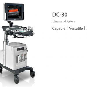
Mindray DC-30 Utrasound Machine
Standard ultrasound systems available today typically allow for a basic diagnosis but lack advanced functionality. Now, with the most competitive price in the industry, the DC-30 is the perfect answer for high quality image performance, with added features such as Auto IMT, iScape, Natural Touch Elastography, UWN Contrast Imaging and Tissue Doppler Imaging.
Performance
PSHTM??(Phase Shift Harmonic Imaging)
Purified Harmonic Imaging for better contrast resolution providing clearer images with excellent resolution and less noise.

iBeamTM
Permits use of multiple scanned angles to form a single image, resulting in enhanced contrast resolution and improved visualization.
iClearTM
Gain improved image quality based on auto structure detection
?? Sharper & Continuous Edges
?? Smooth Uniform Tissues
?? Cleaner ???no echo areas???


Multiple Beam Formation
Maximum 4 times tasking for one transmitted beam, resulting in excellent time resolution and higher frame rate.

Natural Touch Elastography
Based on Mindray???s latest patented technology, natural touch elastography reduces dependence on user operation technique, improving operator???s reproducibility for higher clinical utility.
?? Higher stiffness sensitivity
?? Good stability and reproducibility

iScapeTM
Get a complete and extended view of the anatomical structure through panoramic imaging coupled with velocity indication and forward/backward scan ability making scanning much easier, smoother and more controllable.
UWN (Ultra-Wideband Non-linear) Contrast Imaging
Mindray???s unique contrast imaging technology, utilizing contrast agent characters with both 2nd harmonic and non-linear fundamental signal to get improved S/N ratio for better diagnostic details and longer contrast agent duration for better observation.
ExFOV
Discover better diagnostic information through extended view of the anatomical structure on all convex and linear probes.
B-SteerTM
Your tool for deeper biopsy: allowing adjustments to the scan line to gain better visibility of the needle, nerves and small vessels.

Free Xros MTM
Gain precise anatomical observation by freely placing sample lines at any angle.
TDI
Tissue Doppler Imaging allows you to quantitatively evaluate local myocardial movement and function, providing complete TDI modes for faster and direct diagnoses.

Workflow
iScanHelper
Dedicated inbuilt tutorial software
?? Anatomical diagram illustrations including schematic and ultrasound picture
?? Standard ultrasonogram comparison with real-time scanning
?? Scanning reference picture demonstrating appropriate patient position and probe placement
?? Tips on scanning skills and diagnostic information

Auto IMT
Auto measurement of anterior and posterior wall thickness providing accurate carotid status
Raw Data
Enables optimum flexibility for post processing of the stored images including parameter adjustments, adding comments and measurements, allowing maximum productivity during scanning.
iZoomTM
Gain instant full screen view on the click of a single key.
iTouchTM
Gain instant auto image optimization in B, Color and PW Modes on the click of a single ke.

iStorage
Directly transfer images and reports to PC via network cable.
iStationTM
Mindray???s unique Patient Information Management System allowing you to integrate, review, archive and retrieve patient data effectively.
MedSight
DC-30 lets you transfer clinical images and cine to your IOS or android powered smart device via an interactive app. With MedSight you can now take the clinical examinations with you wherever needed.

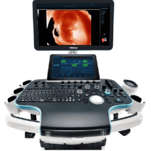
Ergonomics
?? 15''/17?????? LED monitor with easy handle
?? Height adjustable control panel
?? CD/DVD-RW and USB ports
?? Removable transducer holder
?? Built-in battery for live scanning
?? Three active transducer connectors
Clinical Images
Transducers

35C50P

3C5P

6C2P

75L38P

7L4P